This is a SEO version of Journal of Laser Dentistry 1. Click here to view full version
« Previous Page Table of Contents Next Page »Cyst
Mucous cyst of the oral mucosa
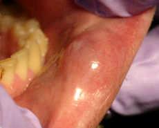
A "mucous cyst of the oral mucosa" (also known as a "mucocele") is a clinical term that refers to two related phenomena: mucus extravasation phenomenon, and mucus retention cyst. The former is a swelling of connective tissue consisting of collected mucin due to a ruptured salivary gland duct usually caused by local trauma, in the case of mucus extravasation phenomenon, and an obstructed salivary duct in the case of a mucus retention cyst. The mucocele is a bluish translucent color, and is more commonly found in children and young adults.
It can be considered a polyp or a cyst.
Locations
The most common location to find a mucocele is the surface of the lower lip. It can also be found on the inner side of the cheek (known as the buccal mucosa), on the anterior ventral tongue, and the floor of the mouth. When found on the floor of the mouth, the mucocele is referred to as a ranula. They are rarely found on the upper lip. As their name suggests they are basically mucus lined cysts
and they can also occur in the Paranasal sinuses most commonly the frontal sinuses, the frontoethomidal region and also in the maxillary sinus. Sphenoid sinus involvement is extremely rare. When the lumen of the vermiform appendix gets blocked due to any factor, again a mucocele can form.
Characteristics
The size of oral mucoceles vary from 1 mm to several centimeters and they usually are slightly transparent with a blue tinge. On palpation, mucoceles may appear fluctuant but can also be firm. Their duration lasts from days to years, and may have recurrent swelling with occasional rupturing of its contents.
Variations
A variant of a mucocele is found on the palate, retromolar pad, and posterior buccal mucosa. Known as a "superficial mucocele", this type presents as single or multiple vesicles and bursts into an ulcer. Despite healing after a few days, superficial mucoceles recur often in the same location.
Histology
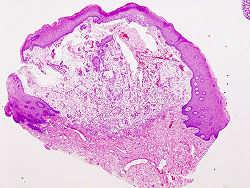
Histopathologic image of extravasation type mucocele of the lower lip. H & E stain.
Microscopically, mucoceles appears as granulation tissue surrounding mucin. Since
This is a SEO version of Journal of Laser Dentistry 1. Click here to view full version
« Previous Page Table of Contents Next Page »