This is a SEO version of Journal of Laser Dentistry 1. Click here to view full version
« Previous Page Table of Contents Next Page »Diagnosis
Diagnosis is usually made by characteristic history and physical examination. Diagnosis can be confirmed by x-ray (80% of salivary gland calculi are visible on x-ray), or by sialogram or ultrasound.
Treatment
Some current treatment options are:
- For small stones, hydration, moist heat, NSAIDs occasionally, and having the patient take any food or beverage that is bitter and/or sour. Sucking on something sour, such as a lemon, may increase salivation and promote spontaneous expulsion of the stone.
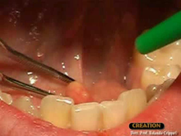
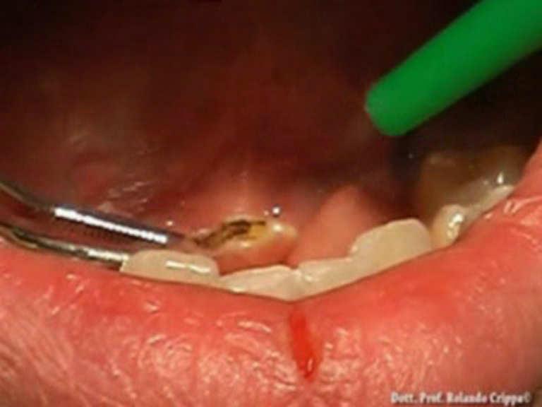
- Some stones may be massaged out by a specialist.
- An ENT or maxillofacial surgeon may canulate the duct to remove the stone (sialotomy).
- A surgeon may make a small incision near the stone to remove it.
- Sialendoscopy
To prevent infection while the stone is lodged in the duct, sometimes antibiotics are used. In some cases when stones continually reoccur the offending salivary duct is removed.
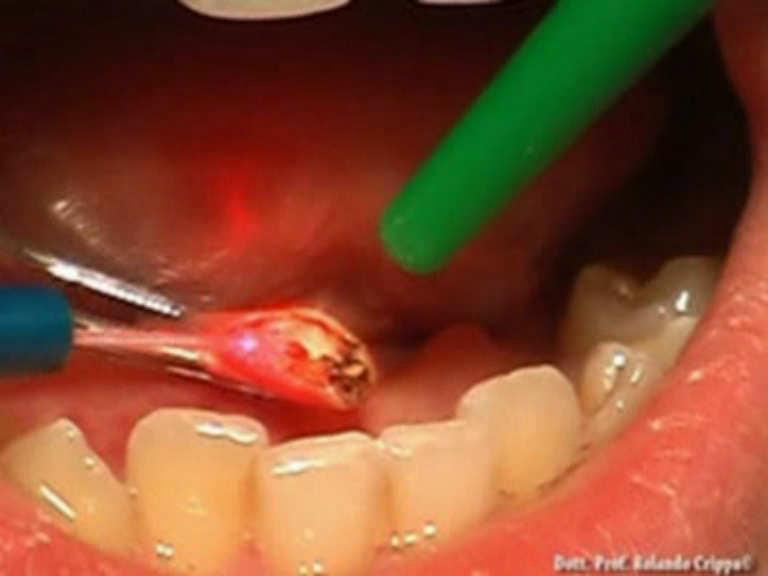
Laser setting
Diode Laser 810nm
Power 2.5 Watt,
pulsed 50 Hz
Time On/Off 10ms
Fibre 400 micron
1
Prof. Dr. Crippa Rolando
This is a SEO version of Journal of Laser Dentistry 1. Click here to view full version
« Previous Page Table of Contents Next Page »