This is a SEO version of Journal of Laser Dentistry 1. Click here to view full version
« Previous Page Table of Contents Next Page »surfaces exhibited signes of laser thermal effect: complete obliteration of dentinal tubles, residual debris and smear layer are still present.
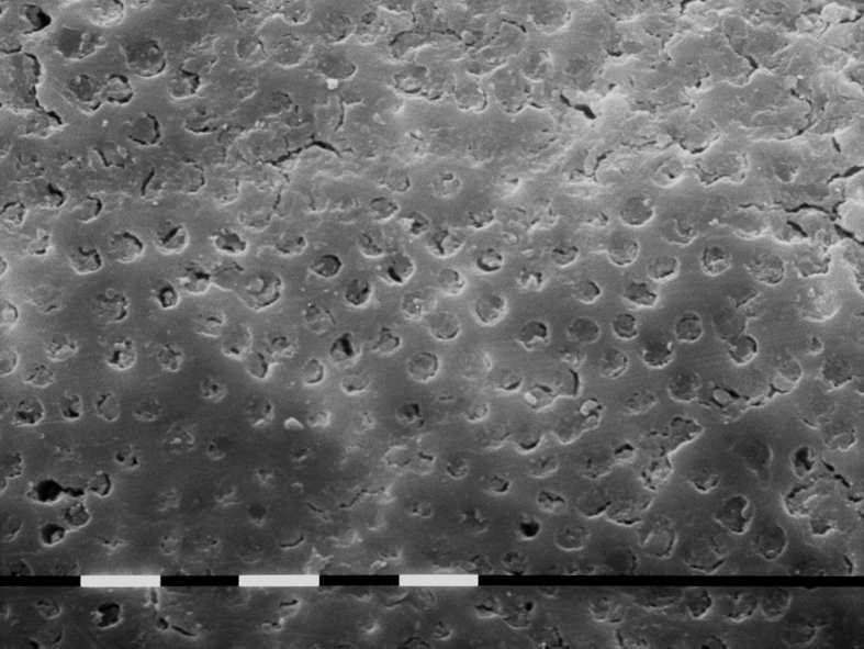
Fig.5 G1 representative sample image at medium third. 810nm diode laser irradiation at 2,5W pulsed mode, 10ms 50%on/off, in EDTA wet canal - 5sec three times: the root canal surfaces are cleaned and exhibit opened dentinal tubules, some residual debris and smear layer still present. No evidence of thermal damage.
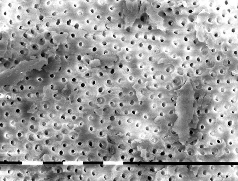
Fig.6 G1 representative sample image at medium third. 810nm diode laser irradiation at 2,5W pulsed mode, 10ms 50%on/off, in EDTA wet canal - 5sec three times: root canal surfaces are enough cleaned, with superficial vaporization of collagen fibers; opened dentinal tubules, some residual debris and smear layer still present.
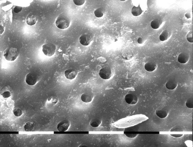
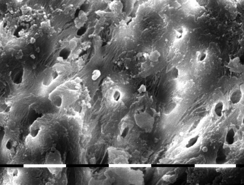
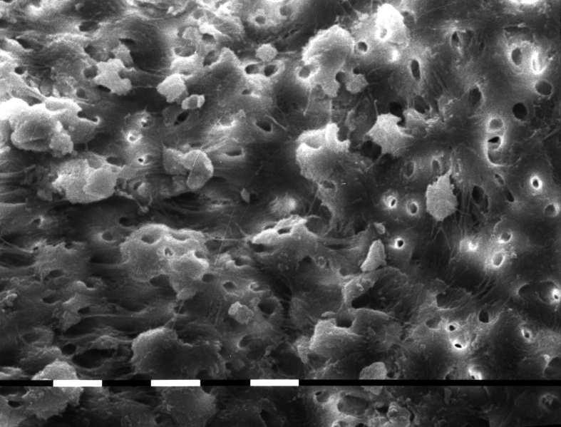
Fig.7-8 G1 representative sample image at medium third. 810nm diode laser irradiation at 2,5W pulsed mode, 10ms 50%on/off, in EDTA wet canal - 5sec three times: root canal surfaces are quite cleaned and opened dentinal tubules, some residual debris and smear layer still present. Evidence of partially maintened organic matrix with exposed and intact collagen fibers both inter and peritubular.
This is a SEO version of Journal of Laser Dentistry 1. Click here to view full version
« Previous Page Table of Contents Next Page »